Skeleton Back Bones Diagram - Anatomy Of The Back Spine And Back Muscles Kenhub - Posted on june 17, 2016 by admin.. C1 is termed the atlas and c2 the axis. The smallest bone in the human body is called the stirrup bone, located deep inside the ear. New users enjoy 60% off. More commonly known as the shoulder blade, the scapula is a flat triangular bone located in the upper back. Find the perfect human back.
Select from premium lower back skeleton of the highest quality. Find the perfect lower back skeleton stock photos and editorial news pictures from getty images. Using this atlas of human anatomy of the spine and back. Spinal vertebrae bone spine vertebra toracica spinal cord spine structure back diagram spine sections spinal cord vertebrae spinal structure health diagram. Download 3,433 human skeleton diagram stock illustrations, vectors & clipart for free or amazingly low rates!
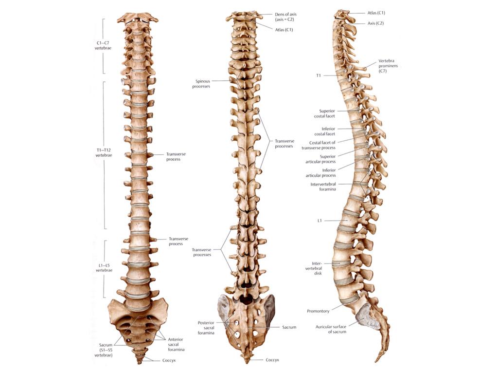
Blank Skeleton Diagram To Label Front And Back Of The Outstanding Skeletal System Anatomy Human Skeletal System Human Skeleton Labeled from i.pinimg.com C1 is termed the atlas and c2 the axis. Bone structure diagram human foot. The cervical spine is further divided into two parts; The bones of the appendicular skeleton provide support and flexibility at the joints and anchor the muscles that move the limbs. Spinal anatomy is a remarkable combination of strong bones, flexible ligaments and tendons, large muscles and highly sensitive nerves. The smallest bone in the human body is called the stirrup bone, located deep inside the ear. Every skeletal muscle has three main parts: Find the perfect lower back skeleton stock photos and editorial news pictures from getty images.
It is composed of 300 bones at birth, but later decreases to 80 bones in the axial skeleton and 126 bones in the appendicular skeleton.
See lumbar spine anatomy diagram stock video clips. Download 3,433 human skeleton diagram stock illustrations, vectors & clipart for free or amazingly low rates! Skeletal diagrams can also be used to show bone development or growth which begins en utero. *the origin, insertion, and belly.* a muscle's origin is where a tendon attaches it to the *less* movable bone. Human body muscles human body organs human body parts human organ diagram body organs diagram anatomy organs anatomy bones heart anatomy body muscle anatomy. Using this atlas of human anatomy of the spine and back. Find the perfect lower back skeleton stock photos and editorial news pictures from getty images. It connects with the collarbone at the front of the body. This article looks at the anatomy of the back, including bones, muscles, and nerves. The upper cervical region (c1 and c2), and the lower cervical region (c3 through c7). There are 358 bones diagram for sale. Hip bones diagram of back and hip bones 9 out of 10 based on 30 ratings. Human backbone diagram, bone, human backbone diagram.
Posted in bones, diagrams | tagged body skeleton, human skeletal anatomy, human skeleton, human. Bones, discs, and joints in your lower back. There are 358 bones diagram for sale. Spinal vertebrae bone spine vertebra toracica spinal cord spine structure back diagram spine sections spinal cord vertebrae spinal structure health diagram. It provides a basic framework in form of skeleton on which everything is else is laid on and anchored to.
The Spine Anatomy Of The Spine Anatomy Medicine Com from anatomy-medicine.com Almost every skeletal muscle works by pulling two or more bones either closer together or further apart. Animals with a skeleton (vertebrates) are a tiny minority as 98 percent of all animals are invertebrates, which means without backbone. osteoporosis is a disease that cases loss of bone tissue. It provides a basic framework in form of skeleton on which everything is else is laid on and anchored to. Every skeletal muscle has three main parts: Your lower back contains 5 vertebral bones stacked above each other with intervertebral discs in between. Select from premium lower back skeleton of the highest quality. This article looks at the anatomy of the back, including bones, muscles, and nerves. The smallest bone in the human body is called the stirrup bone, located deep inside the ear.
New users enjoy 60% off.
Skeletal diagrams can also be used to show bone development or growth which begins en utero. The smallest bone in the human body is called the stirrup bone, located deep inside the ear. Find the perfect human back. Download files and build them with your 3d printer, laser cutter, or cnc. Select from premium lower back skeleton of the highest quality. Find the perfect lower back skeleton stock photos and editorial news pictures from getty images. The red lines point individual bones and the names are writen in singular, the blue lines conect to group of bones and are in plural form. See lumbar spine anatomy diagram stock video clips. Vertebrae, bones, joints, ligaments, muscles, muscular system, fascia, arteries, veins, nerves and various adjacent organs. Every skeletal muscle has three main parts: They support bones, in this case, the vertebrae. Skeleton bone back human body human anchor chart health science diagram. In addition, the broad hip bones provide protection to the delicate internal organs of the pelvis, such as the intestines, urinary bladder, and uterus.
Animals with a skeleton (vertebrates) are a tiny minority as 98 percent of all animals are invertebrates, which means without backbone. osteoporosis is a disease that cases loss of bone tissue. Every skeletal muscle has three main parts: The bones of the skeletal system act as attachment points for the skeletal muscles of the body. Bone diagram forehead (frontal bone) nose bones (nasals) cheek bone (zygoma) upper jaw (maxilla) lower jaw (mandible) breast bone (sternum) upper arm bone. Hip bones diagram of back and hip bones 9 out of 10 based on 30 ratings.
Printable Human Skeleton Diagram Labeled Unlabeled And Blank from i1.wp.com It is designed to be incredibly strong, protecting the highly sensitive nerve roots, yet highly flexible, providing for mobility on many different planes. Almost every skeletal muscle works by pulling two or more bones either closer together or further apart. Important bones diagram human bone anatomy names diagram this arm bones diagram shows all human skeleton diagram of legs human legs bone structure anatomy chart bone anatomy diagram vector illustration of diagram of human bone anatomy royalty free. Human body muscles human body organs human body parts human organ diagram body organs diagram anatomy organs anatomy bones heart anatomy body muscle anatomy. The lumbar spine connects to the thoracic spine above and the hips below. C1 is termed the atlas and c2 the axis. Download 3,433 human skeleton diagram stock illustrations, vectors & clipart for free or amazingly low rates! The bones of the appendicular skeleton provide support and flexibility at the joints and anchor the muscles that move the limbs.
Every skeletal muscle has three main parts:
It is designed to be incredibly strong, protecting the highly sensitive nerve roots, yet highly flexible, providing for mobility on many different planes. Skeletal diagrams can also be used to show bone development or growth which begins en utero. Find the perfect human back. Many muscles that move the trunk and legs, such as our abdominal muscles, attach to the hip bones. On anatomical parts the user can choose to display the various structures in colored illustrations of the anatomy of the back and spine: In addition, the broad hip bones provide protection to the delicate internal organs of the pelvis, such as the intestines, urinary bladder, and uterus. Bone structure diagram human foot. Related posts of human back bones diagram pelvic bone labeled. Human backbone diagram, bone, human backbone diagram. Spinal anatomy is a remarkable combination of strong bones, flexible ligaments and tendons, large muscles and highly sensitive nerves. Bone anatomy of the human body 12 photos of the bone anatomy of the human body back bone structure of human body, bone anatomy of the human body, bone structure in the human body, parts of bones in the human body. Every skeletal muscle has three main parts: Using this atlas of human anatomy of the spine and back.
This article looks at the anatomy of the back, including bones, muscles, and nerves back bones diagram. Posted in bones, diagrams | tagged body skeleton, human skeletal anatomy, human skeleton, human.


0 Komentar